ملف:Mycobacterium tuberculosis 8438 lores.jpg

Mycobacterium_tuberculosis_8438_lores.jpg (700 × 475 بكسل حجم الملف: 49 كيلوبايت، نوع MIME: image/jpeg)

ملخص

الوصف
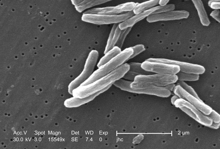
English: Under a high magnification of 15549x, this scanning electron micrograph (SEM) depicted some of the ultrastructural details seen in the cell-wall configuration of a number of Gram-positive Mycobacterium tuberculosis bacteria. As an obligate aerobic organism, M. tuberculosis can only survive in an environment containing oxygen. This bacterium ranges in length between 2-4 microns, with a width of 0.2-0.5 microns. See PHIL 9997 for a colorized version of this image.

TB bacteria become active, and begin to multiply, if the immune system can't stop them from growing. The bacteria attack the body and destroy tissue. If in the lungs, the bacteria can actually create a hole in the lung tissue. Some people develop active TB disease soon after becoming infected, before their immune system can fight off the bacteria. Other people may get sick later, when their immune system becomes weak for another reason.

Babies and young children often have weak immune systems. People infected with HIV, the virus that causes AIDS, have very weak immune systems. Other people can have weak immune systems, too, especially people with any of these conditions: substance abuse; diabetes mellitus; silicosis; cancer of the head or neck; leukemia or Hodgkin's disease; severe kidney disease; low body weight; certain medical treatments (such as corticosteroid treatment or organ transplants); specialized treatment for rheumatoid arthritis, or Crohn's disease.
Français : Mycobacterium tuberculosis grossi 15 549 fois.
Español: Mycobacterium tuberculosis ampliado a 15549x.
中文:掃描電子顯微鏡下的結核桿菌.
Suomi: Mycobacterium tuberculosis 15549-kertaisena suurennoksena.
Čeština: Bakterie Mycobacterium tuberculosis, původce TBC.
한국어: 결핵균의 전자현미경 사진.
Simple English: TB bacteria, shown as 15549x larger by electron microscope.
Kurdî: Girtineke elektronmîkroskobîk a bakteriyên tûberkûlozê pêk tînin.
Afrikaans: 'n Skanderende mikrograaf van Mycobacterium tuberculosis.
粵語: 掃描電子顯微鏡下嘅結核桿菌.
التاريخ
المصدر

ملف الوسائط هذا مَأخُوذٌ مِن مكتبة صُور الصَّحة العامَّة (PHIL) التَّابِعة لمراكز السَّيطرة على الأَمراض والوقاية مِنها ، وله المُعرِّف: #8438.

ملاحظة: ليست ملفَّات مكتبة صُور الصَّحة العامَّة كلُّها في النِّطاق العامِّ، تحقق مِن حالة حقوق التَّأليف والنَّشر ومِن عدم وجود إِلزامٍ بنسب الصورة إلى مُؤَلِّفيها أَو أَصحاب حقوق تأليفها ونشرها.


العربية | Deutsch | English | македонски | slovenščina | +/−

المؤلف
  • Photo Credit: Janice Carr
  • Content Providers(s): CDC/ Dr. Ray Butler; Janice Carr
الترخيص
(إعادة استخدام هذا الملف)
PD-USGov-HHS-CDC
English: This image is in the public domain and thus free of any copyright restrictions. As a matter of courtesy, we request that the content provider be credited and notified in any public or private usage of this image.
إصدارات أخرى

أَعمالٌ مُشتقَّة مِن هذا الملفِّ:  IRG activation following pathogen entry .jpg

ترخيص

Public domain
هذه الصُّورة مِن إِنتاج مراكز السيطرة على الأمراض والوقاية منها (CDC)، التَّابِعة لوزارة الصِّحة والخدمات البشريَّة الأمريكيَّة، التقطت أو أُنشئت بصفتها جزءاً مِن عمل المُوظَفين الرَّسميين فيها. ولأَنَّها عملٌ من إِنتاج الحكومة الاتحادية للولايات المُتحدة الأمريكيَّة، فإِنَّ هذه الصُّورة في النِّطاق العامِّ.

الشروحات

أضف شرحاً من سطر واحد لما يُمثِّله هذا الملف

العناصر المصورة في هذا الملف

يُصوِّر

تاريخ الملف

اضغط على زمن/تاريخ لرؤية الملف كما بدا في هذا الزمن.

زمن/تاريخصورة مصغرةالأبعادمستخدمتعليق
حالي19:45، 18 أبريل 2006تصغير للنسخة بتاريخ 19:45، 18 أبريل 2006700 × 475 (49 كيلوبايت)Patho{{Information| |Description= ID#: 8438 Description: Under a high magnification of 15549x, this scanning electron micrograph (SEM) depicted some of the ultrastructural details seen in the cell wall configuration of a number of Gram-positive Mycobacterium t

الصفحة التالية تستخدم هذا الملف:

الاستخدام العالمي للملف

الويكيات الأخرى التالية تستخدم هذا الملف:

اعرض المزيد من الاستخدام العام لهذا الملف.
بيانات وصفية